
Automating COVID-19 Classification in Chest CT Scans Using Advanced CNN Architectures
DOI:
https://doi.org/10.66279/p8hfyk02Keywords:
Convolutional Networks, COVID-19, CT scan of the lungs, DensNet169, SARS-CoVAbstract
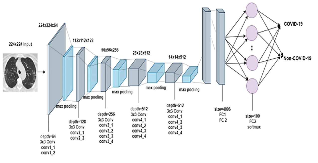
This paper proposes an efficient and fully automated method for classifying COVID-19 using CT scan images of a patient's chest. The study utilizes the publicly available SARS-CoV-2 CT scan dataset, which contains 1252 CT scans positive for SARS-CoV-2 (COVID-19) infection, 1230 CT scans from SARS-CoV-2-negative patients, and a total of 2482 cross-sectional scans. This research explores various topologies designed to enhance the classification accuracy of convolutional neural networks, particularly when dealing with images containing small objects of interest. DenseNet169 is particularly effective in handling small-sized infection patterns commonly observed in COVID-19 cases, as it allows the model to analyze images at varying resolutions effectively without compromising small-object data integrity. Several approaches were evaluated, including VGG19, Xception, ResNet101, DenseNet169, and two custom models: a "custom vanilla" model based on the vanilla architecture and a "custom inception" model based on the inception architecture. Among these, DenseNet169 achieved the highest performance, attaining an impressive accuracy rate of 99.731%.
Downloads
References
[1] F. Song et al., "Emerging 2019 novel coronavirus (2019-nCoV) pneumonia," Radiology, vol. 295, no. 1, pp. 210–217, 2020. DOI: https://doi.org/10.1148/radiol.2020200274
[2] World Health Organization, "Coronavirus disease (COVID-19)," [Online]. Available: https://www.who.int/westernpacific/health-topics/detail/coronavirus. [Accessed: Mar. 20, 2023].
[3] K. G. Andersen et al., "The proximal origin of SARS-CoV-2," Nat. Med., vol. 26, no. 4, pp. 450–452, 2020. DOI: https://doi.org/10.1038/s41591-020-0820-9
[4] E. Dong, H. Du, and L. Gardner, "An interactive web-based dashboard to track COVID-19 in real time," Lancet Infect. Dis., 2020. DOI: https://doi.org/10.1016/S1473-3099(20)30120-1
[5] W. J. Guan et al., "Clinical characteristics of coronavirus disease 2019 in China," N. Engl. J. Med., vol. 382, no. 18, pp. 1708–1720, 2020.
[6] M. Salathé et al., "COVID-19 epidemic in Switzerland: On the importance of testing, contact tracing and isolation," Swiss Med. Wkly., vol. 150, 2020. DOI: https://doi.org/10.4414/smw.2020.20225
[7] S. Cousins, "New Zealand eliminates COVID-19," Lancet, vol. 395, no. 10235, p. 1474, 2020. DOI: https://doi.org/10.1016/S0140-6736(20)31097-7
[8] Z. Hu et al., "Artificial intelligence forecasting of COVID-19 in China," arXiv:2002.07112, 2020. DOI: https://doi.org/10.18562/IJEE.054
[9] Z. Xu et al., "Pathological findings of COVID-19 associated with acute respiratory distress syndrome," Lancet Respir. Med., vol. 8, no. 4, pp. 420–422, 2020. DOI: https://doi.org/10.1016/S2213-2600(20)30076-X
[10] T. Ai et al., "Correlation of chest CT and RT-PCR testing for COVID-19 in China: A report of 1014 cases," Radiology, vol. 296, no. 2, pp. E32–E40, 2020. DOI: https://doi.org/10.1148/radiol.2020200642
[11] American College of Radiology, "ACR recommendations for the use of chest radiography and CT for suspected COVID-19 infection," [Online]. Available: https://www.acr.org. [Accessed: Mar. 20, 2023].
[12] X. Xie et al., "Chest CT for typical coronavirus disease 2019 pneumonia: Relationship to negative RT-PCR testing," Radiology, vol. 296, no. 2, pp. E41–E45, 2020. DOI: https://doi.org/10.1148/radiol.2020200343
[13] E. Soares et al., "A large multiclass dataset of CT scans for COVID-19 identification," Evolving Systems, pp. 1–6, 2023. DOI: https://doi.org/10.1007/s12530-023-09511-2
[14] R. Yamashita et al., "Convolutional neural networks: An overview and application in radiology," Insights Imaging, vol. 9, pp. 611–629, 2018. DOI: https://doi.org/10.1007/s13244-018-0639-9
[15] AIMultiple, "Computer vision in radiology in 2023: Benefits & challenges," [Online]. Available: https://research.aimultiple.com/computer-vision-radiology/. [Accessed: May 20, 2023].
[16] K. He et al., "Identity mappings in deep residual networks," in Proc. ECCV, 2016, pp. 630–645. DOI: https://doi.org/10.1007/978-3-319-46493-0_38
[17] G. Huang et al., "Densely connected convolutional networks," in Proc. CVPR, 2017, pp. 4700–4708.
[18] F. Chollet, "Xception: Deep learning with depthwise separable convolutions," in Proc. CVPR, 2017, pp. 1251–1258. DOI: https://doi.org/10.1109/CVPR.2017.195
[19] K. Simonyan and A. Zisserman, "Very deep convolutional networks for large-scale image recognition," arXiv:1409.1556, 2014.
[20] Towards Data Science, "Review: ResNet — Winner of ILSVRC 2015," [Online]. Available: https://towardsdatascience.com. [Accessed: Aug. 1, 2023].
[21] Towards Data Science, "Understanding and visualizing ResNets," [Online]. Available: https://towardsdatascience.com. [Accessed: Aug. 1, 2023].
[22] Pluralsight, "Introduction to DenseNet with TensorFlow," [Online]. Available: https://www.pluralsight.com. [Accessed: Aug. 1, 2023].
[23] OpenVINO, "DenseNet-169 model," [Online]. Available: https://docs.openvino.ai. [Accessed: Aug. 1, 2023].
[24] J. Deng et al., "ImageNet: A large-scale hierarchical image database," in Proc. CVPR, 2009, pp. 248–255. DOI: https://doi.org/10.1109/CVPR.2009.5206848
[25] F. Chollet, "Keras," 2015.
[26] M. Abadi et al., "TensorFlow: Large-scale machine learning on heterogeneous distributed systems," arXiv:1603.04467, 2016.
[27] L. Li et al., "Artificial intelligence distinguishes COVID-19 from community-acquired pneumonia on chest CT," Radiology, 2020.
[28] M. J. Horry et al., "COVID-19 detection through transfer learning using multimodal imaging data," IEEE Access, vol. 8, pp. 149808–149824, 2020. DOI: https://doi.org/10.1109/ACCESS.2020.3016780
[29] X. Wang et al., "A weakly-supervised framework for COVID-19 classification and lesion localization from chest CT," IEEE Trans. Med. Imaging, vol. 39, no. 8, pp. 2615–2625, 2020. DOI: https://doi.org/10.1109/TMI.2020.2995965
[30] Z. Han et al., "Accurate screening of COVID-19 using attention-based deep 3D multiple instance learning," IEEE Trans. Med. Imaging, vol. 39, no. 8, pp. 2584–2594, 2020. DOI: https://doi.org/10.1109/TMI.2020.2996256
[31] X. Ouyang et al., "Dual-sampling attention network for diagnosis of COVID-19," IEEE Trans. Med. Imaging, vol. 39, no. 8, pp. 2595–2605, 2020. DOI: https://doi.org/10.1109/TMI.2020.2995508
[32] Y. Pathak et al., "Deep bidirectional classification model for COVID-19 patients," IEEE/ACM Trans. Comput. Biol. Bioinform., vol. 18, no. 4, pp. 1234–1241, 2020. DOI: https://doi.org/10.1109/TCBB.2020.3009859
[33] Y. D. Zhang et al., "A five-layer deep CNN with stochastic pooling for COVID-19 diagnosis," Mach. Vis. Appl., vol. 32, pp. 1–13, 2021. DOI: https://doi.org/10.1007/s00138-020-01128-8
[34] A. Waheed et al., "CovidGAN: Data augmentation using auxiliary classifier GAN for improved COVID-19 detection," IEEE Access, vol. 8, pp. 91916–91923, 2020. DOI: https://doi.org/10.1109/ACCESS.2020.2994762
[35] H. A. Owida et al., "Classification of chest X-ray images using wavelet and MFCC features," Eng. Technol. Appl. Sci. Res., vol. 11, no. 4, pp. 7296–7301, 2021. DOI: https://doi.org/10.48084/etasr.4123
Downloads
Published
Data Availability Statement
the data that support the findings of this study are openly available in kaggle at
https://www.kaggle.com/datasets/plameneduardo/sarscov2-ctscan-dataset
Issue
Section
Categories
License
Copyright (c) 2026 Journal of Smart Algorithms and Applications (JSAA)

This work is licensed under a Creative Commons Attribution 4.0 International License.
Journal of Smart Algorithms and Applications (JSAA) content is published under a Creative Commons Attribution License (CCBY). This means that content is freely available to all readers upon publication, and content is published as soon as production is complete.
Journal of Smart Algorithms and Applications (JSAA) seeks to publish the most influential papers that will significantly advance scientific understanding. Selected articles must present new and widely significant data, syntheses, or concepts. They should merit recognition by the wider scientific community and the general public through publication in a reputable scientific journal.