
Certainty-Aware Skin Lesion Segmentation with Post-Hoc Reliability Estimation for the Segment Anything Model
DOI:
https://doi.org/10.66279/hzkw5y24Keywords:
Image Segmentation, Skin Lesion Segmentation, Pixel-wise Certainty Map, Reliability EstimationAbstract
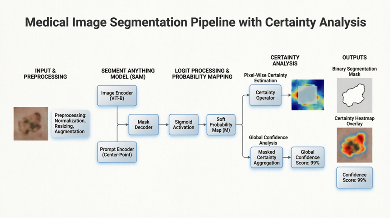
The Segment Anything Model (SAM) represents a major advance in zero-shot visual segmentation, yet it provides purely deterministic outputs without any measure of prediction reliability, a critical limitation for safety-conscious medical imaging applications. This paper introduces a certainty-aware segmentation framework that augments SAM-based zero-shot inference with principled, post-hoc reliability estimation. Three complementary outputs are introduced: a pixel-wise certainty map that identifies spatially localized regions of ambiguity; a global confidence score that provides a scalar measure of overall segmentation trustworthiness; and a quality-flagging mechanism that enables automated screening of unreliable predictions. The framework requires no modification to SAM's architecture and no additional training data, thereby preserving its zero-shot generalization properties. Evaluation on the ISIC 2018 Task 1 skin lesion segmentation benchmark comprising 2,594 dermoscopic images in a fully zero-shot setting yields a mean Dice Similarity Coefficient of 0.820 pm 0.095 and a mean Intersection-over-Union of 0.750 \pm 0.101. A strong positive correlation (Pearson r = 0.84, p < 0.001, n = 2,594) is observed between certainty scores and segmentation quality. High-quality segmentations (DSC> 0.80) are consistently associated with certainty scores above 80%, while low-quality predictions (DSC< 0.70) yield certainty scores below 50%. Stratified analysis confirms a mean DSC difference of over 0.25 between high- and low-certainty tiers (Wilcoxon p < 0.001, Cohen's d = 2.31). These results demonstrate that the proposed certainty metrics reliably track segmentation accuracy and provide a practical mechanism for risk-aware deployment of foundation models in clinical environments.
Downloads
References
[1] A. Kirillov, K. He, R. Girshick, C. Rother, and P. Dollár, “Panoptic segmentation,” in Proceedings of the IEEE/CVF conference on computer vision and pattern recognition, pp. 9404–9413, 2019. DOI: https://doi.org/10.1109/CVPR.2019.00963
[2] M. K. Hasan, L. Dahal, P. N. Samarakoon, F. I. Tushar, and R. Marti, “Dsnet: Automatic dermoscopic skin lesion segmentation,” Computers in biology and medicine, vol. 120, p. 103738, 2020. DOI: https://doi.org/10.1016/j.compbiomed.2020.103738
[3] O. Ronneberger, P. Fischer, and T. Brox, “U-net: Convolutional networks for biomedical image segmentation,” in International Conference on Medical image computing and computer-assisted intervention, pp. 234–241, Springer, 2015. DOI: https://doi.org/10.1007/978-3-319-24574-4_28
[4] M. Z. Alom, C. Yakopcic, M. Hasan, T. M. Taha, and V. K. Asari, “Recurrent residual u-net for medical image segmentation,” Journal of medical imaging, vol. 6, no. 1, pp. 014006–014006, 2019. DOI: https://doi.org/10.1117/1.JMI.6.1.014006
[5] O. Oktay, J. Schlemper, L. L. Folgoc, M. Lee, M. Heinrich, K. Misawa, K. Mori, S. McDonagh, N. Y. Hammerla, B. Kainz, et al., “Attention u-net: Learning where to look for the pancreas,” arXiv preprint arXiv:1804.03999, 2018.
[6] Z. Zhou, M. M. Rahman Siddiquee, N. Tajbakhsh, and J. Liang, “Unet++: A nested u-net architecture for medical image segmentation,” in International workshop on deep learning in medical image analysis, pp. 3–11, Springer, 2018. DOI: https://doi.org/10.1007/978-3-030-00889-5_1
[7] F. Isensee, P. F. Jaeger, S. A. Kohl, J. Petersen, and K. H. Maier-Hein, “nnu-net: a self-configuring method for deep learning-based biomedical image segmentation,” Nature methods, vol. 18, no. 2, pp. 203–211, 2021. DOI: https://doi.org/10.1038/s41592-020-01008-z
[8] R. Bommasani, D. A. Hudson, E. Adeli, R. Altman, S. Arora, S. von Arx, M. S. Bernstein, J. Bohg, A. Bosselut, E. Brunskill, et al., “On the opportunities and risks of foundation models,” arXiv preprint arXiv:2108.07258, 2021.
[9] A. Kirillov, E. Mintun, N. Ravi, H. Mao, C. Rolland, L. Gustafson, T. Xiao, S. Whitehead, A. C. Berg, W.-Y.
Lo, et al., “Segment anything,” in Proceedings of the IEEE/CVF international conference on computer vision, pp. 4015–4026, 2023.
[10] E. Tjoa and C. Guan, “A survey on explainable artificial intelligence (xai): Toward medical xai,” IEEE transactions on neural networks and learning systems, vol. 32, no. 11, pp. 4793–4813, 2020. DOI: https://doi.org/10.1109/TNNLS.2020.3027314
[11] M. Ghassemi, L. Oakden-Rayner, and A. L. Beam, “The false hope of current approaches to explainable artificial intelligence in health care,” The lancet digital health, vol. 3, no. 11, pp. e745–e750, 2021. DOI: https://doi.org/10.1016/S2589-7500(21)00208-9
[12] A. Kendall and Y. Gal, “What uncertainties do we need in bayesian deep learning for computer vision?,” Advances in neural information processing systems, vol. 30, 2017.
[13] B. Lakshminarayanan, A. Pritzel, and C. Blundell, “Simple and scalable predictive uncertainty estimation using deep ensembles,” Advances in neural information processing systems, vol. 30, 2017.
[14] M. Abdar, F. Pourpanah, S. Hussain, D. Rezazadegan, L. Liu, M. Ghavamzadeh, P. Fieguth, X. Cao, A. Khosravi, U. R. Acharya, et al., “A review of uncertainty quantification in deep learning: Techniques, applications and challenges,” Information fusion, vol. 76, pp. 243–297, 2021. DOI: https://doi.org/10.1016/j.inffus.2021.05.008
[15] L. R. Dice, “Measures of the amount of ecologic association between species,” Ecology, vol. 26, no. 3, pp. 297–302, 1945. DOI: https://doi.org/10.2307/1932409
[16] P. Jaccard, “The distribution of the flora in the alpine zone. 1,” New phytologist, vol. 11, no. 2, pp. 37–50, 1912. DOI: https://doi.org/10.1111/j.1469-8137.1912.tb05611.x
[17] Ö. Çiçek, A. Abdulkadir, S. S. Lienkamp, T. Brox, and O. Ronneberger, “3d u-net: learning dense volumetric segmentation from sparse annotation,” in International conference on medical image computing and computer-assisted intervention, pp. 424–432, Springer, 2016. DOI: https://doi.org/10.1007/978-3-319-46723-8_49
[18] L.-C. Chen, Y. Zhu, G. Papandreou, F. Schroff, and H. Adam, “Encoder-decoder with atrous separable convolution for semantic image segmentation,” in Proceedings of the European conference on computer vision (ECCV), pp. 801–818, 2018. DOI: https://doi.org/10.1007/978-3-030-01234-2_49
[19] A. Mehrtash, W. M. Wells, C. M. Tempany, P. Abolmaesumi, and T. Kapur, “Confidence calibration and predictive uncertainty estimation for deep medical image segmentation,” IEEE transactions on medical imaging, vol. 39, no. 12, pp. 3868–3878, 2020. DOI: https://doi.org/10.1109/TMI.2020.3006437
[20] A. Dosovitskiy, L. Beyer, A. Kolesnikov, D. Weissenborn, X. Zhai, T. Unterthiner, M. Dehghani, M. Minderer, G. Heigold, S. Gelly, et al., “An image is worth 16x16 words: Transformers for image recognition at scale,” arXiv preprint arXiv:2010.11929, 2020.
[21] J. Ma, Y. He, F. Li, L. Han, C. You, and B. Wang, “Segment anything in medical images,” Nature communications, vol. 15, no. 1, p. 654, 2024. DOI: https://doi.org/10.1038/s41467-024-44824-z
[22] J. Cheng, J. Ye, Z. Deng, J. Chen, T. Li, H. Wang, Y. Su, Z. Huang, J. Chen, L. Jiang, et al., “Sam-med2d,” arXiv preprint arXiv:2308.16184, 2023.
[23] N. Ravi, V. Gabeur, Y.-T. Hu, R. Hu, C. Ryali, T. Ma, H. Khedr, R. Rädle, C. Rolland, L. Gustafson, et al., “Sam 2: Segment anything in images and videos,” arXiv preprint arXiv:2408.00714, 2024.
[24] K. Zhang and D. Liu, “Customized segment anything model for medical image segmentation,” arXiv preprint arXiv:2304.13785, 2023. DOI: https://doi.org/10.2139/ssrn.4495221
[25] N. Codella, V. Rotemberg, P. Tschandl, M. E. Celebi, S. Dusza, D. Gutman, B. Helba, A. Kalloo, K. Liopyris, M. Marchetti, et al., “Skin lesion analysis toward melanoma detection 2018: A challenge hosted by the international skin imaging collaboration (isic),” arXiv preprint arXiv:1902.03368, 2019.
Downloads
Published
Data Availability Statement
The ISIC 2018 Task 1 dataset is publicly available from the International Skin Imaging Collaboration (ISIC) archive: https://challenge.isic-archive.com/data/#2018. The SAM pretrained checkpoint (sam_vit_b_01ec64.pth) is available at https://github.com/facebookresearch/segment-anything.
Issue
Section
Categories
License
Copyright (c) 2026 Journal of Smart Algorithms and Applications (JSAA)

This work is licensed under a Creative Commons Attribution 4.0 International License.
Journal of Smart Algorithms and Applications (JSAA) content is published under a Creative Commons Attribution License (CCBY). This means that content is freely available to all readers upon publication, and content is published as soon as production is complete.
Journal of Smart Algorithms and Applications (JSAA) seeks to publish the most influential papers that will significantly advance scientific understanding. Selected articles must present new and widely significant data, syntheses, or concepts. They should merit recognition by the wider scientific community and the general public through publication in a reputable scientific journal.