
Artificial Intelligence for Automated Semen Analysis from Microscopy to Clinical Translation
DOI:
https://doi.org/10.66279/p63yh388Keywords:
Artificial Intelligence, Automated Semen Analysis, Clinical Translation, Hybrid Systems, Deep LearningAbstract
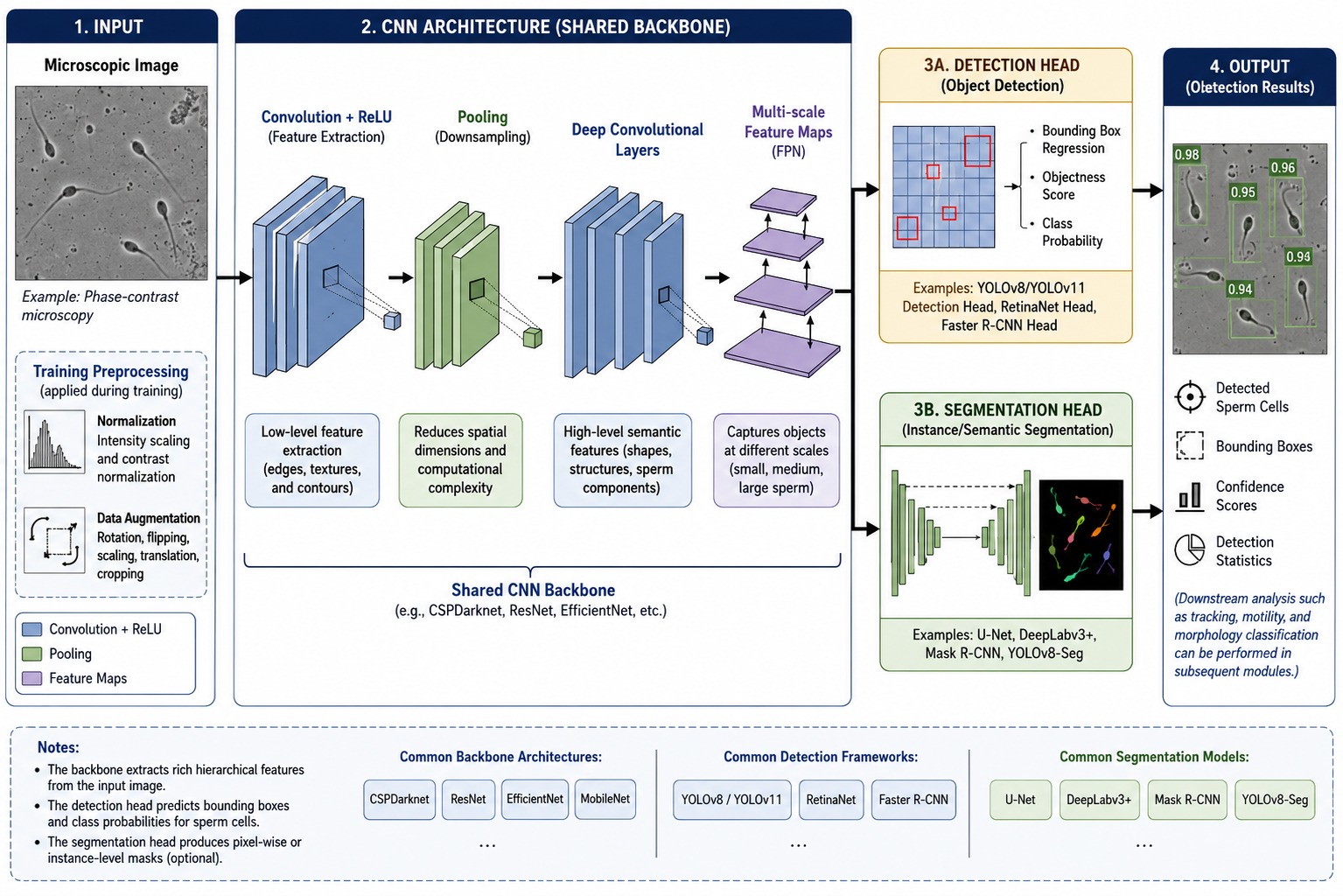
Male-factor infertility is commonly evaluated using semen analysis; however, manual assessment of sperm concentration, motility, and morphology remains susceptible to observer variability and laboratory heterogeneity. This review synthesizes artificial intelligence methods for automated semen analysis, covering sperm detection, segmentation, morphology classification, motility assessment, tracking, and hybrid computational pipelines. A literature search was conducted across PubMed/MEDLINE, IEEE Xplore, Scopus, Web of Science Core Collection, and Google Scholar. Eligible studies were peer-reviewed, English-language investigations that applied machine-learning or deep-learning methods to at least one semen-analysis task and reported quantitative performance measures. The reviewed studies demonstrate recurring use of transfer learning, one-stage object detection, U-Net- and Mask R-CNN-based segmentation, video-level convolutional models, transformer-based architectures, model ensembles, and generative data augmentation. Although many studies report high performance on curated internal datasets, comparisons across studies are limited by differences in species, imaging modality, annotation procedures, evaluation metrics, and data-partitioning strategies. External validation, patient- or donor-level leakage control, calibration, uncertainty estimation, and prospective assessment of clinical utility remain uncommon. Hybrid systems appear most justified when their components address complementary failure modes and when their
incremental value is evaluated against an appropriately matched single-model baseline; however, such comparisons are not consistently reported. Overall, the available evidence supports the technical feasibility of artificial intelligence for automated semen analysis but does not yet establish readiness for routine clinical deployment. Translation will require harmonized multicenter datasets, consensus-based annotation, leakage-aware external validation, task-specific reporting standards, defined human oversight, and regulatory planning appropriate to the intended use and jurisdiction.
Downloads
References
[1] A. Agarwal, A. Mulgund, A. Hamada, and M. R. Chyatte, “A unique view on male infertility around the globe,” Reproductive biology and endocrinology, vol. 13, no. 1, p. 37, 2015. DOI: https://doi.org/10.1186/s12958-015-0032-1
[2] W. H. Organization et al., WHO laboratory manual for the examination and processing of human semen. World Health Organization, 2021.
[3] K. Siddharth, T. Kumar, and M. Zabihullah, “Interobserver variability in semen analysis: findings from a quality control initiative,” Cureus, vol. 15, no. 10, 2023. DOI: https://doi.org/10.7759/cureus.46388
[4] S. T. Mortimer, G. Van der Horst, and D. Mortimer, “The future of computer-aided sperm analysis,” Asian journal of andrology, vol. 17, no. 4, p. 545, 2015. DOI: https://doi.org/10.4103/1008-682X.154312
[5] J.-w. Choi, L. Alkhoury, L. F. Urbano, P. Masson, M. VerMilyea, and M. Kam, “An assessment tool for computer-assisted semen analysis (casa) algorithms,” Scientific reports, vol. 12, no. 1, p. 16830, 2022. DOI: https://doi.org/10.1038/s41598-022-20943-9
[6] G. Litjens, T. Kooi, B. E. Bejnordi, A. A. A. Setio, F. Ciompi, M. Ghafoorian, J. A. Van Der Laak, B. Van Ginneken, and C. I. Sánchez, “A survey on deep learning in medical image analysis,” Medical image analysis, vol. 42, pp. 60–88, 2017. DOI: https://doi.org/10.1016/j.media.2017.07.005
[7] T. Sato, H. Kishi, S. Murakata, Y. Hayashi, T. Hattori, S. Nakazawa, Y. Mori, M. Hidaka, Y. Kasahara, A. Kusuhara, et al., “A new deep-learning model using yolov3 to support sperm selection during intracytoplasmic sperm injection procedure,” Reproductive Medicine and Biology, vol. 21, no. 1, p. e12454, 2022. DOI: https://doi.org/10.1002/rmb2.12454
[8] F. Shaker, S. A. Monadjemi, J. Alirezaie, and A. R. Naghsh-Nilchi, “A dictionary learning approach for human sperm heads classification,” Computers in biology and medicine, vol. 91, pp. 181–190, 2017. DOI: https://doi.org/10.1016/j.compbiomed.2017.10.009
[9] J. Riordon, C. McCallum, and D. Sinton, “Deep learning for the classification of human sperm,” Computers in biology and medicine, vol. 111, p. 103342, 2019. DOI: https://doi.org/10.1016/j.compbiomed.2019.103342
[10] H. O. Ilhan and N. Aydin, “Smartphone based sperm counting-an alternative way to the visual assessment technique in sperm concentration analysis,” Multimedia Tools and Applications, vol. 79, no. 9, pp. 6409–6435, 2020. DOI: https://doi.org/10.1007/s11042-019-08421-3
[11] S. Javadi and S. A. Mirroshandel, “A novel deep learning method for automatic assessment of human sperm images,” Computers in biology and medicine, vol. 109, pp. 182–194, 2019. DOI: https://doi.org/10.1016/j.compbiomed.2019.04.030
[12] T. B. Haugen, O. Witczak, S. A. Hicks, L. Björndahl, J. M. Andersen, and M. A. Riegler, “Sperm motility assessed by deep convolutional neural networks into who categories,” Scientific Reports, vol. 13, no. 1, p. 14777, 2023. DOI: https://doi.org/10.1038/s41598-023-41871-2
[13] J. Wang, Y. Jin, A. Jiang, W. Chen, G. Shan, Y. Gu, Y. Ming, J. Li, C. Yue, Z. Huang, et al., “Testing the generalizability and effectiveness of deep learning models among clinics: sperm detection as a pilot study,” Reproductive Biology and Endocrinology, vol. 22, no. 1, p. 59, 2024. DOI: https://doi.org/10.1186/s12958-024-01232-8
[14] V. Valiuškait ˙e, V. Raudonis, R. Maskeli ¯unas, R. Damaševičius, and T. Krilavičius, “Deep learning based evaluation of spermatozoid motility for artificial insemination,” Sensors, vol. 21, no. 1, 2021. DOI: https://doi.org/10.3390/s21010072
[15] L. Spencer, J. Fernando, F. Akbaridoust, K. Ackermann, and R. Nosrati, “Ensembled deep learning for the classification of human sperm head morphology,” Advanced Intelligent Systems, vol. 4, no. 10, p. 2200111, 2022. DOI: https://doi.org/10.1002/aisy.202200111
[16] S. Zou, C. Li, H. Sun, P. Xu, J. Zhang, P. Ma, Y. Yao, X. Huang, and M. Grzegorzek, “Tod-cnn: An effective convolutional neural network for tiny object detection in sperm videos,” Computers in Biology and Medicine, vol. 146, p. 105543, 2022. DOI: https://doi.org/10.1016/j.compbiomed.2022.105543
[17] D. Wu, O. Badamjav, V. Reddy, M. Eisenberg, and B. Behr, “A preliminary study of sperm identification in microdissection testicular sperm extraction samples with deep convolutional neural networks,” Asian Journal of Andrology, vol. 23, pp. 135–139, 10 2020. DOI: https://doi.org/10.4103/aja.aja_66_20
[18] A. Keller, M. Maus, E. Keller, and K. Kerns, “Deep learning classification method for boar sperm morphology analysis,” Andrology, vol. 13, no. 6, pp. 1615–1625, 2025. DOI: https://doi.org/10.1111/andr.13758
[19] R. Marín and V. Chang, “Impact of transfer learning for human sperm segmentation using deep learning,” Computers in Biology and Medicine, vol. 136, p. 104687, 2021. DOI: https://doi.org/10.1016/j.compbiomed.2021.104687
[20] P. Lei, M. Saadat, M. G. Hassani, and C. Shu, “Deep learning models for multi-part morphological segmentation and evaluation of live unstained human sperm,” Sensors, vol. 25, no. 10, p. 3093, 2025. DOI: https://doi.org/10.3390/s25103093
[21] T. B. Haugen, S. A. Hicks, J. M. Andersen, O. Witczak, H. L. Hammer, R. Borgli, P. Halvorsen, and M. Riegler, “Visem: A multimodal video dataset of human spermatozoa,” in Proceedings of the 10th ACM Multimedia Systems Conference, pp. 261–266, 2019. DOI: https://doi.org/10.1145/3304109.3325814
[22] C. Zhang, Y. Zhang, Z. Chang, and C. Li, “Sperm yolov8e-trackevd: A novel approach for sperm detection and tracking,” Sensors, vol. 24, no. 11, 2024. DOI: https://doi.org/10.3390/s24113493
[23] P. Hidayatullah, X. Wang, T. Yamasaki, T. L. Mengko, R. Munir, A. Barlian, E. Sukmawati, and S. Supraptono, “Deepsperm: A robust and real-time bull sperm-cell detection in densely populated semen videos,” Computer Methods and Programs in Biomedicine, vol. 209, p. 106302, 2021. DOI: https://doi.org/10.1016/j.cmpb.2021.106302
[24] M. Yuzkat, H. O. Ilhan, and N. Aydin, “Detection of sperm cells by single-stage and two-stage deep object detectors,” Biomedical Signal Processing and Control, vol. 83, p. 104630, 2023. DOI: https://doi.org/10.1016/j.bspc.2023.104630
[25] R. Zhu, Y. Cui, J. Huang, E. Hou, J. Zhao, Z. Zhou, and H. Li, “Yolov5s-sa: light-weighted and improved yolov5s for sperm detection,” Diagnostics, vol. 13, no. 6, p. 1100, 2023. DOI: https://doi.org/10.3390/diagnostics13061100
[26] M. Dobrovolny, J. Benes, J. Langer, O. Krejcar, and A. Selamat, “Study on sperm-cell detection using yolov5 architecture with labaled dataset,” Genes, vol. 14, no. 2, p. 451, 2023. DOI: https://doi.org/10.3390/genes14020451
[27] R. Liu, M. Wang, M. Wang, J. Yin, Y. Yuan, and J. Liu, “Automatic microscopy analysis with transfer learning for classification of human sperm,” Applied Sciences, vol. 11, no. 12, p. 5369, 2021. DOI: https://doi.org/10.3390/app11125369
[28] M. I. Mahali, J.-S. Leu, J. T. Darmawan, C. Avian, N. Bachroin, S. W. Prakosa, M. Faisal, and N. A. S. Putro, “A dual
architecture fusion and autoencoder for automatic morphological classification of human sperm,” Sensors, vol. 23, no. 14, p. 6613, 2023. DOI: https://doi.org/10.3390/s23146613
[29] B. Cansiz, H. O. Ilhan, and G. Serbes, “Loss-based ensemble generative adversarial network model for enhancing the
sperm morphology classification,” Advanced Intelligent Systems, vol. 8, no. 2, p. e202500441, 2026.
[30] D. Somasundaram and M. Nirmala, “Faster region convolutional neural network and semen tracking algorithm for sperm analysis,” Computer Methods and Programs in Biomedicine, vol. 200, p. 105918, 2021. DOI: https://doi.org/10.1016/j.cmpb.2020.105918
[31] J. Redmon, S. Divvala, R. Girshick, and A. Farhadi, “You only look once: Unified, real-time object detection,” in Proceedings of the IEEE conference on computer vision and pattern recognition, pp. 779–788, 2016. DOI: https://doi.org/10.1109/CVPR.2016.91
[32] W. Liu, D. Anguelov, D. Erhan, C. Szegedy, S. Reed, C.-Y. Fu, and A. C. Berg, “Ssd: Single shot multibox detector,” in European conference on computer vision, pp. 21–37, Springer, 2016. DOI: https://doi.org/10.1007/978-3-319-46448-0_2
[33] N. Carion, F. Massa, G. Synnaeve, N. Usunier, A. Kirillov, and S. Zagoruyko, “End-to-end object detection with transformers,” in European conference on computer vision, pp. 213–229, Springer, 2020. DOI: https://doi.org/10.1007/978-3-030-58452-8_13
[34] X. Zhu, W. Su, L. Lu, B. Li, X. Wang, and J. Dai, “Deformable detr: Deformable transformers for end-to-end object detection,” arXiv preprint arXiv:2010.04159, 2020.
[35] T. Prangemeier, C. Reich, and H. Koeppl, “Attention-based transformers for instance segmentation of cells in microstructures,” in 2020 IEEE international conference on Bioinformatics and Biomedicine (BIBM), pp. 700–707, IEEE, 2020. DOI: https://doi.org/10.1109/BIBM49941.2020.9313305
[36] Y. Zhao, W. Lv, S. Xu, J. Wei, G. Wang, Q. Dang, Y. Liu, and J. Chen, “Detrs beat yolos on real-time object detection,” in 2024 IEEE/CVF Conference on Computer Vision and Pattern Recognition (CVPR), pp. 16965–16974, IEEE, 2024. DOI: https://doi.org/10.1109/CVPR52733.2024.01605
[37] O. Ronneberger, P. Fischer, and T. Brox, “U-net: Convolutional networks for biomedical image segmentation,” in International Conference on Medical image computing and computer-assisted intervention, pp. 234–241, Springer, 2015. DOI: https://doi.org/10.1007/978-3-319-24574-4_28
[38] R. A. Movahed, E. Mohammadi, and M. Orooji, “Automatic segmentation of sperm’s parts in microscopic images of human semen smears using concatenated learning approaches,” Computers in Biology and Medicine, vol. 109, pp. 242–253, 2019. DOI: https://doi.org/10.1016/j.compbiomed.2019.04.032
[39] H. Kaiming, G. Georgia, D. Piotr, and G.-s. Ross, “Mask r-cnn,” in Proceedings of the IEEE international conference on computer vision, vol. 2017, pp. 2961–2969, 2017.
[40] Q. Lv, X. Yuan, J. Qian, X. Li, H. Zhang, and S. Zhan, “An improved u-net for human sperm head segmentation,” Neural Processing Letters, vol. 54, no. 1, pp. 537–557, 2022. DOI: https://doi.org/10.1007/s11063-021-10643-2
[41] J. Chen, Y. Lu, Q. Yu, X. Luo, E. Adeli, Y. Wang, L. Lu, A. L. Yuille, and Y. Zhou, “Transunet: Transformers make strong encoders for medical image segmentation,” arXiv preprint arXiv:2102.04306, 2021.
[42] N. Otsu, “A threshold selection method from gray-level histograms,” IEEE transactions on systems, man, and cybernetics, vol. 9, no. 1, pp. 62–66, 1979. DOI: https://doi.org/10.1109/TSMC.1979.4310076
[43] J. Sauvola and M. Pietikäinen, “Adaptive document image binarization,” Pattern recognition, vol. 33, no. 2, pp. 225–236, 2000. DOI: https://doi.org/10.1016/S0031-3203(99)00055-2
[44] S. Beucher, “The watershed transformation applied to image segmentation,” Scanning microscopy, vol. 1992, no. 6, p. 28, 1992.
[45] M. Kass, A. Witkin, and D. Terzopoulos, “Snakes: Active contour models,” International journal of computer vision, vol. 1, no. 4, pp. 321–331, 1988. DOI: https://doi.org/10.1007/BF00133570
[46] S. Osher and J. A. Sethian, “Fronts propagating with curvature-dependent speed: Algorithms based on hamilton-jacobi formulations,” Journal of computational physics, vol. 79, no. 1, pp. 12–49, 1988. DOI: https://doi.org/10.1016/0021-9991(88)90002-2
[47] I. Iqbal, G. Mustafa, and J. Ma, “Deep learning-based morphological classification of human sperm heads,” Diagnostics, vol. 10, no. 5, p. 325, 2020. DOI: https://doi.org/10.3390/diagnostics10050325
[48] P. Hernández-Herrera, V. Abonza, J. Sanchez-Contreras, A. Darszon, and A. Guerrero, “Deep learning-based classification and segmentation of sperm head and flagellum for image-based flow cytometry,” Computación y Sistemas, vol. 27, no. 4, pp. 1133–1145, 2023. DOI: https://doi.org/10.13053/cys-27-4-4772
[49] S. J. Pan and Q. Yang, “A survey on transfer learning,” IEEE Transactions on knowledge and data engineering, vol. 22, no. 10, pp. 1345–1359, 2009. DOI: https://doi.org/10.1109/TKDE.2009.191
[50] A. Abbasi, E. Miahi, and S. A. Mirroshandel, “Effect of deep transfer and multi-task learning on sperm abnormality detection,” Computers in Biology and Medicine, vol. 128, p. 104121, 2021. DOI: https://doi.org/10.1016/j.compbiomed.2020.104121
[51] H. O. Ilhan and G. Serbes, “Sperm morphology analysis by using the fusion of two-stage fine-tuned deep networks,” Biomedical Signal Processing and Control, vol. 71, p. 103246, 2022. DOI: https://doi.org/10.1016/j.bspc.2021.103246
[52] M. Dobrovolny, J. Benes, O. Krejcar, and A. Selamat, “Sperm-cell detection using yolov5 architecture,” in International Work-Conference on Bioinformatics and Biomedical Engineering, pp. 319–330, Springer, 2022. DOI: https://doi.org/10.1007/978-3-031-07802-6_27
[53] S. Shahzad, M. Ilyas, M. I. U. Lali, H. T. Rauf, S. Kadry, and E. A. Nasr, “Sperm abnormality detection using sequential deep neural network,” Mathematics, vol. 11, no. 3, p. 515, 2023. DOI: https://doi.org/10.3390/math11030515
[54] L. Prabaharan and N. Saravanan, “A three stage framework for abnormality detection in sperm cell images using cnn,”
Biomedical Signal Processing and Control, vol. 99, p. 106827, 2025. DOI: https://doi.org/10.1016/j.bspc.2024.106827
[55] S. Shahali, M. Murshed, L. Spencer, O. Tunc, L. Pisarevski, J. Conceicao, R. McLachlan, M. K. O’Bryan, K. Ackermann, D. Zander-Fox, et al., “Morphology classification of live unstained human sperm using ensemble deep learning,” Advanced Intelligent Systems, vol. 6, no. 11, p. 2400141, 2024. DOI: https://doi.org/10.1002/aisy.202400141
[56] A. Aristoteles, A. Syarif, S. Sutyarso, and F. R. Lumbanraja, “Identification of human sperm based on morphology
using the you only look once version 4 algorithm,” International Journal of Advanced Computer Science and Applications, vol. 13, no. 7, pp. 424–431, 2022.
[57] Z. Yin, T. Kanade, and M. Chen, “Understanding the phase contrast optics to restore artifact-free microscopy images for segmentation,” Medical image analysis, vol. 16, no. 5, pp. 1047–1062, 2012. DOI: https://doi.org/10.1016/j.media.2011.12.006
[58] K. Chatzimeletiou, A. Fleva, T.-T. Nikolopoulos, M. Markopoulou, G. Zervakakou, K. Papanikolaou, G. Anifandis, A. Gianakou, and G. Grimbizis, “Evaluation of sperm dna fragmentation using two different methods: Tunel via fluorescence microscopy, and flow cytometry,” Medicina, vol. 59, no. 7, p. 1313, 2023. DOI: https://doi.org/10.3390/medicina59071313
[59] L. Noy, I. Barnea, S. K. Mirsky, D. Kamber, M. Levi, and N. T. Shaked, “Sperm-cell dna fragmentation prediction using label-free quantitative phase imaging and deep learning,” Cytometry Part A, vol. 103, no. 6, pp. 470–478, 2023. DOI: https://doi.org/10.1002/cyto.a.24703
[60] A. Agarwal, C.-L. Cho, S. C. Esteves, and A. Majzoub, “Reactive oxygen species and sperm dna fragmentation,”
Translational andrology and urology, vol. 6, no. Suppl 4, p. S695, 2017. DOI: https://doi.org/10.21037/tau.2017.05.40
[61] K. Zuiderveld, “Contrast limited adaptive histogram equalization,” in Graphics Gems IV (P. S. Heckbert, ed.), pp. 474–485, San Diego, CA, USA: Academic Press, 1994. DOI: https://doi.org/10.1016/B978-0-12-336156-1.50061-6
[62] R. C. Gonzalez and R. E. Woods, Digital Image Processing. Upper Saddle River, NJ, USA: Prentice Hall, 2 ed., 2002.
[63] K. Zhang, W. Zuo, Y. Chen, D. Meng, and L. Zhang, “Beyond a gaussian denoiser: Residual learning of deep cnn for image denoising,” IEEE transactions on image processing, vol. 26, no. 7, pp. 3142–3155, 2017. DOI: https://doi.org/10.1109/TIP.2017.2662206
[64] T. Ojala, M. Pietikainen, and T. Maenpaa, “Multiresolution gray-scale and rotation invariant texture classification with local binary patterns,” IEEE Transactions on pattern analysis and machine intelligence, vol. 24, no. 7, pp. 971–987, 2002. DOI: https://doi.org/10.1109/TPAMI.2002.1017623
[65] J. G. Daugman, “Complete discrete 2-d gabor transforms by neural networks for image analysis and compression,”
IEEE Transactions on acoustics, speech, and signal processing, vol. 36, no. 7, pp. 1169–1179, 1988. DOI: https://doi.org/10.1109/29.1644
[66] H. O. Ilhan, I. O. Sigirci, G. Serbes, and N. Aydin, “A fully automated hybrid human sperm detection and classification system based on mobile-net and the performance comparison with conventional methods,” Medical & biological engineering & computing, vol. 58, no. 5, pp. 1047–1068, 2020. DOI: https://doi.org/10.1007/s11517-019-02101-y
[67] S. G. Goodson, S. White, A. M. Stevans, S. Bhat, C.-Y. Kao, S. Jaworski, T. R. Marlowe, M. Kohlmeier, L. McMillan,
S. H. Zeisel, et al., “Casanova: a multiclass support vector machine model for the classification of human sperm
motility patterns,” Biology of reproduction, vol. 97, no. 5, pp. 698–708, 2017. DOI: https://doi.org/10.1093/biolre/iox120
[68] I. J. Goodfellow, J. Pouget-Abadie, M. Mirza, B. Xu, D. Warde-Farley, S. Ozair, A. Courville, and Y. Bengio, “Generative adversarial nets,” Advances in neural information processing systems, vol. 27, 2014.
[69] M. E. Kandel, M. Fanous, C. Best-Popescu, and G. Popescu, “Real-time halo correction in phase contrast imaging,” Biomedical optics express, vol. 9, no. 2, pp. 623–635, 2018. DOI: https://doi.org/10.1364/BOE.9.000623
[70] E. Lewandowska, D. Węsierski, M. Mazur-Milecka, J. Liss, and A. Jezierska, “Ensembling noisy segmentation masks of blurred sperm images,” Computers in Biology and Medicine, vol. 166, p. 107520, 2023. DOI: https://doi.org/10.1016/j.compbiomed.2023.107520
[71] A. Chen, J. Zhang, M. M. Rahaman, H. Sun, T. Zeng, M. Grzegorzek, F.-L. Fan, C. Li, et al., “Active: A deep model for sperm and impurity detection in microscopic videos,” arXiv preprint arXiv:2301.06002, 2023. DOI: https://doi.org/10.1109/BIBM62325.2024.10822549
[72] M. E. Kandel, M. Rubessa, Y. R. He, S. Schreiber, S. Meyers, L. Matter Naves, M. K. Sermersheim, G. S. Sell, M. J. Szewczyk, N. Sobh, et al., “Reproductive outcomes predicted by phase imaging with computational specificity of spermatozoon ultrastructure,” Proceedings of the National Academy of Sciences, vol. 117, no. 31, pp. 18302–18309, 2020. DOI: https://doi.org/10.1073/pnas.2001754117
[73] F. Shaker, “Human sperm head morphology dataset (hushem),” Mendeley Data, vol. 3, p. 2018, 2018.
[74] V. Chang, A. Garcia, N. Hitschfeld, and S. Härtel, “Gold-standard for computer-assisted morphological sperm analysis,” Computers in biology and medicine, vol. 83, pp. 143–150, 2017. DOI: https://doi.org/10.1016/j.compbiomed.2017.03.004
[75] A. Chen, C. Li, S. Zou, M. M. Rahaman, Y. Yao, H. Chen, H. Yang, P. Zhao, W. Hu, W. Liu, et al., “Svia dataset: a new dataset of microscopic videos and images for computer-aided sperm analysis,” Biocybernetics and Biomedical Engineering, vol. 42, no. 1, pp. 204–214, 2022. DOI: https://doi.org/10.1016/j.bbe.2021.12.010
[76] H. O. Ilhan and N. Aydin, “A novel data acquisition and analyzing approach to spermiogram tests,” Biomedical Signal Processing and Control, vol. 41, pp. 129–139, 2018. DOI: https://doi.org/10.1016/j.bspc.2017.11.009
[77] V. Dubey, D. Popova, A. Ahmad, G. Acharya, P. Basnet, D. S. Mehta, and B. S. Ahluwalia, “Partially spatially coherent digital holographic microscopy and machine learning for quantitative analysis of human spermatozoa under oxidative stress condition,” Scientific reports, vol. 9, no. 1, p. 3564, 2019. DOI: https://doi.org/10.1038/s41598-019-39523-5
[78] M. L. D. Garcia, D. A. P. Soto, and L. S. Mihaylova, “A bag of features based approach for classification of motile sperm cells,” in 2017 IEEE International Conference on Internet of Things (iThings) and IEEE Green Computing and Communications (GreenCom) and IEEE Cyber, Physical and Social Computing (CPSCom) and IEEE Smart Data (SmartData), pp. 104–109, IEEE, 2017. DOI: https://doi.org/10.1109/iThings-GreenCom-CPSCom-SmartData.2017.21
[79] M. S. Nissen, O. Krause, K. Almstrup, S. Kjærulff, T. T. Nielsen, and M. Nielsen, “Convolutional neural networks for segmentation and object detection of human semen,” in Scandinavian conference on image analysis, pp. 397–406, Springer, 2017. DOI: https://doi.org/10.1007/978-3-319-59126-1_33
[80] S. K. Mirsky, I. Barnea, M. Levi, H. Greenspan, and N. T. Shaked, “Automated analysis of individual sperm cells using stain-free interferometric phase microscopy and machine learning,” Cytometry Part A, vol. 91, no. 9, pp. 893–900, 2017. DOI: https://doi.org/10.1002/cyto.a.23189
[81] A. Butola, D. Popova, D. K. Prasad, A. Ahmad, A. Habib, J. C. Tinguely, P. Basnet, G. Acharya, P. Senthilkumaran,
D. S. Mehta, et al., “High spatially sensitive quantitative phase imaging assisted with deep neural network for classification of human spermatozoa under stressed condition,” Scientific reports, vol. 10, no. 1, p. 13118, 2020. DOI: https://doi.org/10.1038/s41598-020-69857-4
[82] A. Bijar, M. Mikaeili, R. Khayati, et al., “Fully automatic identification and discrimination of sperm’s parts in microscopic images of stained human semen smear,” Journal of Biomedical Science and Engineering, vol. 5, no. 2012, 2012. DOI: https://doi.org/10.4236/jbise.2012.57049
[83] K.-K. Tseng, Y. Li, C.-Y. Hsu, H.-N. Huang, M. Zhao, and M. Ding, “Computer-assisted system with multiple feature fused support vector machine for sperm morphology diagnosis,” BioMed research international, vol. 2013, no. 1, p. 687607, 2013. DOI: https://doi.org/10.1155/2013/687607
[84] F. Shaker, S. A. Monadjemi, and A. R. Naghsh-Nilchi, “Automatic detection and segmentation of sperm head, acrosome and nucleus in microscopic images of human semen smears,” Computer methods and programs in biomedicine, vol. 132, pp. 11–20, 2016. DOI: https://doi.org/10.1016/j.cmpb.2016.04.026
Downloads
Published
Data Availability Statement
DATA AVAILABILITY
No new primary clinical datasets were generated during this study. This work is based on a structured review and
comparative analysis of previously published literature.
Issue
Section
Categories
License
Copyright (c) 2026 The Authors

This work is licensed under a Creative Commons Attribution 4.0 International License.
Journal of Smart Algorithms and Applications (JSAA) seeks to publish the most influential papers that will significantly advance scientific understanding. Selected articles must present new and widely significant data, syntheses, or concepts. They should merit recognition by the wider scientific community and the general public through publication in a reputable scientific journal.
Journal of Smart Algorithms and Applications (JSAA) content is published under a Creative Commons Attribution 4.0 International (CC BY 4.0) License. This means that content is freely available to all readers upon publication, and content is published as soon as production is complete.
JSAA Journal
SIRG Publisher