
A Comprehensive Review of Artificial Intelligence for Lumbar Spine MRI Analysis and Clinical Assessment
DOI:
https://doi.org/10.66279/r8h6j935Keywords:
Artificial Intelligence, Lumbar Spine MRI, Clinical Foundations, Deep Learning, Intervertebral Disc DegenerationAbstract
Lower back pain is one of the major diseases to disability worldwide, with lumbar intervertebral disc degeneration (IVDD), disc herniation (DH), and lumbar spinal stenosis (LSS) being the most common underlying causes. Magnetic resonance imaging (MRI) is the imaging modality of choice for assessing these conditions; however, conventional MRI interpretation continues to be time-consuming, subjective, and prone to considerable interobserver variability. Recent advances in artificial intelligence (AI), particularly deep learning and advanced AI frameworks, have shown strong potential for optimizing the accuracy, consistency, and efficiency of lumbar spine MRI analysis. This review offers an in-depth overview of AI applications for lumbar spine MRI
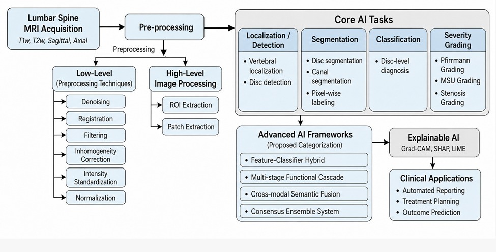
analysis of IVDD, DH and LSS. It includes lumbar spine anatomy, pathophysiology of diseases, MRI grading systems and publicly available datasets including SPIDER, LumbarDISC and LSpineSMRI. AI methodologies are comprehensively reviewed across deep learning methods and advanced AI frameworks, covering feature–classifier hybrids, multi-stage functional cascades, cross-modal semantic fusion, and consensus ensemble systems, alongside widely adopted architectures such as CNNs, U-Net, YOLO, and transformer-based models for localization, segmentation, classification, and severity grading. Reported performance in the reviewed studies showed Dice similarity coefficients exceeding 0.90 for segmentation tasks and AUC values up to approximately 0.98 for lumbar spinal stenosis assessment. The review further discusses the key challenges to clinical translation, encompassing dataset heterogeneity, class imbalance, domain shift, limited external validation, and insufficient interpretability, while outlining future research priorities such as multimodal learning, self-supervised
learning, foundation models, and explainable AI (XAI) to enhance model robustness, transparency, and clinical applicability. This review presents a clinically driven and technically structured perspective on the current landscape of AI for lumbar spine MRI analysis and identifies directions for developing more robust, interpretable, and clinically deployable diagnostic systems.
Downloads
References
[1] A. Raja’S, J. J. Corso, V. Chaudhary, and G. Dhillon, “Automatic diagnosis of lumbar disc herniation with shape and appearance features from mri,” in Medical Imaging 2010: Computer-Aided Diagnosis, vol. 7624, pp. 364–372, SPIE, 2010. DOI: https://doi.org/10.1117/12.842199
[2] J. N. Katz and M. B. Harris, “Lumbar spinal stenosis,” New England Journal of Medicine, vol. 358, no. 8, pp. 818–825, 2008. DOI: https://doi.org/10.1056/NEJMcp0708097
[3] D. F. Fardon, A. L. Williams, E. J. Dohring, F. R. Murtagh, S. L. G. Rothman, and G. K. Sze, “Lumbar disc nomenclature: version 2.0: Recommendations of the combined task forces of the north american spine society, the american society of spine radiology and the american society of neuroradiology,” The spine journal, vol. 14, no. 11, pp. 2525–2545, 2014. DOI: https://doi.org/10.1016/j.spinee.2014.04.022
[4] V. Tumko, J. Kim, N. Uspenskaia, S. Honig, F. Abel, D. R. Lebl, I. Hotalen, S. Kolisnyk, M. Kochnev, A. Rusakov, et al., “A neural network model for detection and classification of lumbar spinal stenosis on mri,” European Spine Journal, vol. 33, no. 3, pp. 941–948, 2024. DOI: https://doi.org/10.1007/s00586-023-08089-2
[5] R. U. Din, X. Cheng, and H. Yang, “Diagnostic role of magnetic resonance imaging in low back pain caused by vertebral endplate degeneration,” Journal of Magnetic Resonance Imaging, vol. 55, no. 3, pp. 755–771, 2022. DOI: https://doi.org/10.1002/jmri.27858
[6] V. P. Grover, J. M. Tognarelli, M. M. Crossey, I. J. Cox, S. D. Taylor-Robinson, and M. J. McPhail, “Magnetic resonance imaging: principles and techniques: lessons for clinicians,” Journal of clinical and experimental hepatology, vol. 5, no. 3, pp. 246–255, 2015. DOI: https://doi.org/10.1016/j.jceh.2015.08.001
[7] J. A. Carrino, J. D. Lurie, A. N. Tosteson, T. D. Tosteson, E. J. Carragee, J. Kaiser, M. R. Grove, E. Blood, L. H. Pearson, J. N. Weinstein, et al., “Lumbar spine: reliability of MR imaging findings,” Radiology, vol. 250, no. 1, pp. 161–170, 2009. DOI: https://doi.org/10.1148/radiol.2493071999
[8] J. Lin, H. Zhang, and H. Shang, “Convolutional neural network incorporating multiple attention mechanisms for mri classification of lumbar spinal stenosis,” Bioengineering, vol. 11, no. 10, p. 1021, 2024. DOI: https://doi.org/10.3390/bioengineering11101021
[9] M. Khalifa and M. Albadawy, “Ai in diagnostic imaging: revolutionising accuracy and efficiency,” Computer Methods and programs in biomedicine update, vol. 5, p. 100146, 2024. DOI: https://doi.org/10.1016/j.cmpbup.2024.100146
[10] N. C. Lehnen, R. Haase, J. Faber, T. Rüber, H. Vatter, A. Radbruch, and F. C. Schmeel, “Detection of degenerative changes on mr images of the lumbar spine with a convolutional neural network: a feasibility study,” Diagnostics, vol. 11, no. 5, p. 902, 2021. DOI: https://doi.org/10.3390/diagnostics11050902
[11] M. Alabsi, A. AQashlan, R. Mohamed, A. Taha, et al., “Automated brain tumor segmentation via yolov8-derived spatial prompts for the segment anything model,” Computational Discovery and Intelligent Systems (CDIS), vol. 3, no. 2, pp. 150–163, 2026. DOI: https://doi.org/10.66279/vv8n5p41
[12] R. Compte, I. Granville Smith, A. Isaac, N. Danckert, T. McSweeney, P. Liantis, and F. M. Williams, “Are current machine learning applications comparable to radiologist classification of degenerate and herniated discs and modic change? a systematic review and meta-analysis,” European Spine Journal, vol. 32, no. 11, pp. 3764–3787, 2023. DOI: https://doi.org/10.1007/s00586-023-07718-0
[13] M. Hussain, D. Koundal, and J. Manhas, “Deep learning-based diagnosis of disc degenerative diseases using mri: a comprehensive review,” Computers and Electrical Engineering, vol. 105, p. 108524, 2023. DOI: https://doi.org/10.1016/j.compeleceng.2022.108524
[14] M. A. Adams and P. J. Roughley, “What is intervertebral disc degeneration, and what causes it?,” Spine, vol. 31, no. 18, pp. 2151–2161, 2006. DOI: https://doi.org/10.1097/01.brs.0000231761.73859.2c
[15] W. Mbarki, M. Bouchouicha, S. Frizzi, F. Tshibasu, L. B. Farhat, and M. Sayadi, “Lumbar spine discs classification based on deep convolutional neural networks using axial view mri,” Interdisciplinary Neurosurgery, vol. 22, p. 100837, 2020. DOI: https://doi.org/10.1016/j.inat.2020.100837
[16] J. Dowdell, M. Erwin, T. Choma, A. Vaccaro, J. Iatridis, and S. K. Cho, “Intervertebral disk degeneration and repair,” Neurosurgery, vol. 80, no. 3S, pp. S46–S54, 2017. DOI: https://doi.org/10.1093/neuros/nyw078
[17] S. Salamat, J. Hutchings, C. Kwong, J. Magnussen, and M. J. Hancock, “The relationship between quantitative measures of disc height and disc signal intensity with pfirrmann score of disc degeneration,” Springerplus, vol. 5, no. 1, p. 829, 2016. DOI: https://doi.org/10.1186/s40064-016-2542-5
[18] C. W. Pfirrmann, A. Metzdorf, M. Zanetti, J. Hodler, and N. Boos, “Magnetic resonance classification of lumbar intervertebral disc degeneration,” spine, vol. 26, no. 17, pp. 1873–1878, 2001. DOI: https://doi.org/10.1097/00007632-200109010-00011
[19] A. N. Laiwalla, A. Ratnaparkhi, D. Zarrin, K. Cook, I. Li, B. Wilson, T. Florence, B. Yoo, B. Salehi, B. Gaonkar, et al., “Lumbar spinal canal segmentation in cases with lumbar stenosis using deep-u-net ensembles,” World Neurosurgery, vol. 178, pp. e135–e140, 2023. DOI: https://doi.org/10.1016/j.wneu.2023.07.009
[20] E. Ebrahimzadeh, F. Fayaz, F. Ahmadi, and M. Nikravan, “A machine learning-based method in order to diagnose lumbar disc herniation disease by mr image processing,” MedLife Open Access, vol. 1, no. 1, p. 1, 2018.
[21] G. Valarmathi and S. N. Devi, “Automatic localization and classification of intervertebral disc herniation using hybrid classifier,” Biomedical Signal Processing and Control, vol. 86, p. 105291, 2023. DOI: https://doi.org/10.1016/j.bspc.2023.105291
[22] J. N. Katz, Z. E. Zimmerman, H. Mass, and M. C. Makhni, “Diagnosis and management of lumbar spinal stenosis: a review,” Jama, vol. 327, no. 17, pp. 1688–1699, 2022. DOI: https://doi.org/10.1001/jama.2022.5921
[23] D. S. Kreiner, W. O. Shaffer, J. L. Baisden, T. J. Gilbert, J. T. Summers, J. F. Toton, S. W. Hwang, R. C. Mendel, and C. A. Reitman, “An evidence-based clinical guideline for the diagnosis and treatment of degenerative lumbar spinal stenosis (update),” The Spine Journal, vol. 13, no. 7, pp. 734–743, 2013. DOI: https://doi.org/10.1016/j.spinee.2012.11.059
[24] F. Galbusera, G. Casaroli, and T. Bassani, “Artificial intelligence and machine learning in spine research,” JOR spine, vol. 2, no. 1, p. e1044, 2019. DOI: https://doi.org/10.1002/jsp2.1044
[25] S. Sudirman, A. Al Kafri, F. Natalia, H. Meidia, N. Afriliana, W. Al-Rashdan, M. Bashtawi, and M. Al-Jumaily, “Lumbar spine mri dataset,” 2019.
[26] R. F. Masood, T. Hassan, M. Akram, I. Taj, M. Qureshi, and M. Khan, “Composite dataset of lumbar spine mid-sagittal images with annotations and clinically relevant spinal measurements,” Mendeley Data: London, UK, 2021.
[27] F. Natalia, S. Sudirman, D. Ruslim, and A. Al-Kafri, “Lumbar spine mri annotation with intervertebral disc height and pfirrmann grade predictions,” PLoS One, vol. 19, no. 5, p. e0302067, 2024. DOI: https://doi.org/10.1371/journal.pone.0302067
[28] J. W. van der Graaf, M. L. van Hooff, C. F. Buckens, M. Rutten, J. L. van Susante, R. J. Kroeze, M. de Kleuver, B. van Ginneken, and N. Lessmann, “Lumbar spine segmentation in mr images: a dataset and a public benchmark,” Scientific Data, vol. 11, no. 1, p. 264, 2024. DOI: https://doi.org/10.1038/s41597-024-03090-w
[29] T. J. Richards, A. E. Flanders, E. Colak, L. M. Prevedello, R. L. Ball, F. Kitamura, J. Mongan, M. Vazirabad, H.-M. Lin, A. Kendell, et al., “The rsna lumbar degenerative imaging spine classification (lumbardisc) dataset,” Radiology: Artificial Intelligence, vol. 8, no. 2, p. e250480, 2026.
[30] N. Delfan, M. Parvaresh, M. Shafieeha, and B. Moshiri, “Lspinesmri: A comprehensive dataset of non-contrast lumbar spine stenosis mri examinations,” 2025.
[31] B. Zárate-Kalfópulos, F. Reyes-Tarrago, L. A. Navarro-Aceves, C. L. García-Ramos, A. A. Reyes-Sánchez, A. Alpízar-Aguirre, and L. M. Rosales-Olivarez, “Characteristics of spinopelvic sagittal alignment in lumbar degenerative disease,” World neurosurgery, vol. 126, pp. e417–e421, 2019. DOI: https://doi.org/10.1016/j.wneu.2019.02.067
[32] F. D’Antoni, F. Russo, L. Ambrosio, L. Bacco, L. Vollero, G. Vadalà, M. Merone, R. Papalia, and V. Denaro, “Artificial intelligence and computer aided diagnosis in chronic low back pain: A systematic review,” International journal of environmental research and public health, vol. 19, no. 10, p. 5971, 2022. DOI: https://doi.org/10.3390/ijerph19105971
[33] A. Wang, C. Zou, S. Yuan, N. Fan, P. Du, T. Wang, and L. Zang, “Deep learning assisted segmentation of the lumbar intervertebral disc: a systematic review and meta-analysis,” Journal of Orthopaedic Surgery and Research, vol. 19, no. 1, p. 496, 2024. DOI: https://doi.org/10.1186/s13018-024-05002-5
[34] T. Wang, R. Chen, N. Fan, L. Zang, S. Yuan, P. Du, Q. Wu, A. Wang, J. Li, X. Kong, et al., “Machine learning and deep learning for diagnosis of lumbar spinal stenosis: systematic review and meta-analysis,” Journal of medical Internet research, vol. 26, p. e54676, 2024. DOI: https://doi.org/10.2196/54676
[35] W. Liawrungrueang, J.-B. Park, W. Cholamjiak, P. Sarasombath, and K. D. Riew, “Artificial intelligence-assisted mri diagnosis in lumbar degenerative disc disease: a systematic review,” Global Spine Journal, vol. 15, no. 2, pp. 1405–1418, 2025. DOI: https://doi.org/10.1177/21925682241274372
[36] P. Zhao and S. Zhu, “Advances and challenges in ai-assisted mri for lumbar disc degeneration detection and classification,” European Spine Journal, vol. 35, no. 3, pp. 1291–1300, 2026. DOI: https://doi.org/10.1007/s00586-025-09179-z
[37] X. Wei, L. Gengwu, C. Chao, L. Yifan, S. Shang, H. Ruixi, J. Yunhan, Z. Xiaodong, and L. Zhikun, “Correlations between the sagittal plane parameters of the spine and pelvis and lumbar disc degeneration,” Journal of orthopaedic surgery and research, vol. 13, no. 1, p. 137, 2018. DOI: https://doi.org/10.1186/s13018-018-0838-6
[38] J. F. Griffith, Y.-X. J. Wang, G. E. Antonio, K. C. Choi, A. Yu, A. T. Ahuja, and P. C. Leung, “Modified pfirrmann grading system for lumbar intervertebral disc degeneration,” Spine, vol. 32, no. 24, pp. E708–E712, 2007. DOI: https://doi.org/10.1097/BRS.0b013e31815a59a0
[39] Z. Soydan, E. Bayramoglu, D. U. Urut, A. C. Iplikcioglu, and C. Sen, “Tracing the disc: the novel qualitative morphometric mri based disc degeneration classification system,” JOR spine, vol. 7, no. 1, p. e1321, 2024. DOI: https://doi.org/10.1002/jsp2.1321
[40] L. W. Mysliwiec, J. Cholewicki, M. D. Winkelpleck, and G. P. Eis, “Msu classification for herniated lumbar discs on mri: toward developing objective criteria for surgical selection,” European spine journal, vol. 19, no. 7, pp. 1087–1093, 2010. DOI: https://doi.org/10.1007/s00586-009-1274-4
[41] R. I. Riesenburger, M. G. Safain, R. Ogbuji, J. Hayes, and S. W. Hwang, “A novel classification system of lumbar disc degeneration,” Journal of Clinical Neuroscience, vol. 22, no. 2, pp. 346–351, 2015. DOI: https://doi.org/10.1016/j.jocn.2014.05.052
[42] C. Aprill and N. Bogduk, “High-intensity zone: a diagnostic sign of painful lumbar disc on magnetic resonance imaging,” The British journal of radiology, vol. 65, no. 773, pp. 361–369, 1992. DOI: https://doi.org/10.1259/0007-1285-65-773-361
[43] M. A. Yoon, S.-J. Hong, C. H. Kang, K.-S. Ahn, and B. H. Kim, “T1rho and t2 mapping of lumbar intervertebral disc: correlation with degeneration and morphologic changes in different disc regions,” Magnetic resonance imaging, vol. 34, no. 7, pp. 932–939, 2016. DOI: https://doi.org/10.1016/j.mri.2016.04.024
[44] O. Togao, A. Hiwatashi, T. Wada, K. Yamashita, K. Kikuchi, C. Tokunaga, J. Keupp, M. Yoneyama, and H. Honda, “A qualitative and quantitative correlation study of lumbar intervertebral disc degeneration using glycosaminoglycan chemical exchange saturation transfer, pfirrmann grade, and t1-????,” American Journal of Neuroradiology, vol. 39, no. 7, pp. 1369–1375, 2018. DOI: https://doi.org/10.3174/ajnr.A5657
[45] W. Yi, J. Zhao, W. Tang, H. Yin, L. Yu, Y. Wang, and W. Tian, “Deep learning-based high-accuracy detection for lumbar and cervical degenerative disease on t2 weighted mr images,” European Spine Journal, vol. 32, no. 11, pp. 3807–3814, 2023. DOI: https://doi.org/10.1007/s00586-023-07641-4
[46] Y. LeCun, Y. Bengio, and G. Hinton, “Deep learning,” nature, vol. 521, no. 7553, pp. 436–444, 2015. DOI: https://doi.org/10.1038/nature14539
[47] A. Piórkowski, R. Obuchowicz, A. Urbanik, and M. Strzelecki, “Advances in musculoskeletal imaging and their applications,” Journal of Clinical Medicine, vol. 12, no. 20, 2023. DOI: https://doi.org/10.3390/jcm12206585
[48] R. Obuchowicz, M. Strzelecki, and A. Piórkowski, “Clinical applications of artificial intelligence in medical imaging and image processing—a review,” Cancers, vol. 16, no. 10, 2024. DOI: https://doi.org/10.3390/cancers16101870
[49] S. Pang, C. Pang, L. Zhao, Y. Chen, Z. Su, Y. Zhou, M. Huang, W. Yang, H. Lu, and Q. Feng, “Spineparsenet: spine parsing for volumetric mr image by a two-stage segmentation framework with semantic image representation,” IEEETransactions on Medical Imaging, vol. 40, no. 1, pp. 262–273, 2020. DOI: https://doi.org/10.1109/TMI.2020.3025087
[50] H.-D. Zheng, Y.-L. Sun, D.-W. Kong, M.-C. Yin, J. Chen, Y.-P. Lin, X.-F. Ma, H.-S. Wang, G.-J. Yuan, M. Yao,
et al., “Deep learning-based high-accuracy quantitation for lumbar intervertebral disc degeneration from mri,” Nature communications, vol. 13, no. 1, p. 841, 2022. DOI: https://doi.org/10.1038/s41467-022-28387-5
[51] N. T. Singh, C. Kaur, A. Chaudhary, and S. Goyal, “Preprocessing of medical images using deep learning: A
comprehensive review,” in 2023 Second International Conference on Augmented Intelligence and Sustainable Systems(ICAISS), pp. 521–527, IEEE, 2023.
[52] P. Chlap, H. Min, N. Vandenberg, J. Dowling, L. Holloway, and A. Haworth, “A review of medical image dataaugmentation techniques for deep learning applications,” Journal of medical imaging and radiation oncology, vol. 65, no. 5, pp. 545–563, 2021. DOI: https://doi.org/10.1111/1754-9485.13261
[53] R. Girshick, J. Donahue, T. Darrell, and J. Malik, “Rich feature hierarchies for accurate object detection and semantic segmentation,” in Proceedings of the IEEE conference on computer vision and pattern recognition, pp. 580–587, DOI: https://doi.org/10.1109/CVPR.2014.81
2014.
[54] S. Pang, Z. Su, S. Leung, I. B. Nachum, B. Chen, Q. Feng, and S. Li, “Direct automated quantitative measurement of spine by cascade amplifier regression network with manifold regularization,” Medical image analysis, vol. 55, DOI: https://doi.org/10.1016/j.media.2019.04.012
pp. 103–115, 2019.
[55] T. Martín-Noguerol, M. O. Miranda, T. J. Amrhein, F. Paulano-Godino, P. Xiberta, J. C. Vilanova, and A. Luna, “The role of artificial intelligence in the assessment of the spine and spinal cord,” European journal of radiology, vol. 161, p. 110726, 2023. DOI: https://doi.org/10.1016/j.ejrad.2023.110726
[56] E. Tsalera, A. Papadakis, M. Samarakou, and I. Voyiatzis, “Feature extraction with handcrafted methods and convolutional neural networks for facial emotion recognition,” Applied Sciences, vol. 12, no. 17, p. 8455, 2022. DOI: https://doi.org/10.3390/app12178455
[57] R. Ponnusamy, S. Sathyamoorthy, and K. Manikandan, “A review of image classification approaches and techniques,” International Journal of Recent Trends in Engineering & Research, vol. 3, no. 3, pp. 1–5, 2017. DOI: https://doi.org/10.23883/IJRTER.2017.3033.XTS7Z
[58] I. Guyon and A. Elisseeff, “An introduction to variable and feature selection,” Journal of machine learning research, vol. 3, no. Mar, pp. 1157–1182, 2003.
[59] B. Han and L. S. Davis, “Density-based multifeature background subtraction with support vector machine,” IEEE Transactions on Pattern Analysis and Machine Intelligence, vol. 34, no. 5, pp. 1017–1023, 2011. DOI: https://doi.org/10.1109/TPAMI.2011.243
[60] J.-T. Lu, S. Pedemonte, B. Bizzo, S. Doyle, K. P. Andriole, M. H. Michalski, R. G. Gonzalez, and S. R. Pomerantz, “Deep spine: automated lumbar vertebral segmentation, disc-level designation, and spinal stenosis grading using deep learning,” in Machine learning for healthcare conference, pp. 403–419, PMLR, 2018.
[61] Q. Pan, K. Zhang, L. He, Z. Dong, L. Zhang, X. Wu, Y. Wu, and Y. Gao, “Automatically diagnosing disk bulge and disk herniation with lumbar magnetic resonance images by using deep convolutional neural networks: method development study,” JMIR medical informatics, vol. 9, no. 5, p. e14755, 2021. DOI: https://doi.org/10.2196/14755
[62] F. Niemeyer, F. Galbusera, Y. Tao, A. Kienle, M. Beer, and H.-J. Wilke, “A deep learning model for the accurate and reliable classification of disc degeneration based on mri data,” Investigative radiology, vol. 56, no. 2, pp. 78–85, 2021. DOI: https://doi.org/10.1097/RLI.0000000000000709
[63] J.-Y. Tsai, I. Y.-J. Hung, Y. L. Guo, Y.-K. Jan, C.-Y. Lin, T. T.-F. Shih, B.-B. Chen, and C.-W. Lung, “Lumbar disc herniation automatic detection in magnetic resonance imaging based on deep learning,” Frontiers in Bioengineering and Biotechnology, vol. 9, p. 708137, 2021. DOI: https://doi.org/10.3389/fbioe.2021.708137
[64] W. Liawrungrueang, P. Kim, V. Kotheeranurak, K. Jitpakdee, and P. Sarasombath, “Automatic detection, classification, and grading of lumbar intervertebral disc degeneration using an artificial neural network model,” Diagnostics, vol. 13, no. 4, p. 663, 2023. DOI: https://doi.org/10.3390/diagnostics13040663
[65] T. Šušteršič, V. Ranković, V. Milovanović, V. Kovačević, L. Rasulić, and N. Filipović, “A deep learning model for automatic detection and classification of disc herniation in magnetic resonance images,” IEEE Journal of Biomedical and Health Informatics, vol. 26, no. 12, pp. 6036–6046, 2022. DOI: https://doi.org/10.1109/JBHI.2022.3209585
[66] T. Shahzadi, M. U. Ali, F. Majeed, M. U. Sana, R. M. Diaz, M. A. Samad, and I. Ashraf, “Nerve root compression analysis to find lumbar spine stenosis on mri using cnn,” Diagnostics, vol. 13, no. 18, p. 2975, 2023. DOI: https://doi.org/10.3390/diagnostics13182975
[67] J. Qian, G. Su, X. Shu, K. Shen, B. Chen, and X. Wang, “Lumbar disc herniation diagnosis using deep learning on mri,” Journal of Radiation Research and Applied Sciences, vol. 17, no. 3, p. 100988, 2024. DOI: https://doi.org/10.1016/j.jrras.2024.100988
[68] K. Chen, L. Zheng, H. Zhao, and Z. Wang, “Deep learning-based intelligent diagnosis of lumbar diseases with multi-angle view of intervertebral disc,” Mathematics, vol. 12, no. 13, p. 2062, 2024. DOI: https://doi.org/10.3390/math12132062
[69] Y. Xu, S. Zheng, Q. Tian, Z. Kou, W. Li, X. Xie, and X. Wu, “Deep learning model for grading and localization of lumbar disc herniation on magnetic resonance imaging,” Journal of Magnetic Resonance Imaging, vol. 61, no. 1, pp. 364–375, 2025. DOI: https://doi.org/10.1002/jmri.29403
[70] D. Pan, Y.-X. Pan, H. Wang, Q.-J. Liu, and C.-M. Qiu, “Nearest-neighbor dual-path contrastive learning for lumbar disc herniation mri image classification,” IEEE Access, vol. 13, pp. 92906–92920, 2025. DOI: https://doi.org/10.1109/ACCESS.2025.3571014
[71] M. Abed, Z. Al-Qaysi, and M. Suzani, “Improving lumbar disc bulging detection in mri spinal imaging: A deep learning approach,” Al-Salam Journal for Engineering and Technology, vol. 4, no. 1, pp. 1–19, 2025. DOI: https://doi.org/10.55145/ajest.2025.04.01.001
[72] M. A. Sayed, A. Rahman, S. M. Nasif, S. Halder, A. Hossain, H. Ahmed, and M. A. Kadir, “Automated lumbar intervertebral disc identification and herniation detection in mr images using cascade cnn architecture,” Informatics in Medicine Unlocked, vol. 55, p. 101648, 2025. DOI: https://doi.org/10.1016/j.imu.2025.101648
[73] M. Ebrahim, M. Alsmirat, and M. Al-Ayyoub, “Advanced disk herniation computer aided diagnosis system,” Scientific
Reports, vol. 14, no. 1, p. 8071, 2024.
[74] Y. Sarı and N. Aydın Atasoy, “Enhanced disc herniation classification using grey wolf optimization based on hybrid feature extraction and deep learning methods,” Tomography, vol. 11, no. 1, p. 1, 2024. DOI: https://doi.org/10.3390/tomography11010001
[75] D. Zhang, J. Du, J. Shi, Y. Zhang, S. Jia, X. Liu, Y. Wu, Y. An, S. Zhu, D. Pan, et al., “A fully automatic mri-guided decision support system for lumbar disc herniation using machine learning,” JOR spine, vol. 7, no. 2, p. e1342, 2024. DOI: https://doi.org/10.1002/jsp2.1342
[76] R. Dong, X. Cheng, M. Kang, and Y. Qu, “Classification of lumbar spine disorders using large language models and mri segmentation,” BMC Medical Informatics and Decision Making, vol. 24, no. 1, p. 343, 2024. DOI: https://doi.org/10.1186/s12911-024-02740-8
[77] M. A. Sayed, G. M. Rahman, M. S. Islam, M. A. Islam, J. Park, H. Ahmed, A. Hossain, and R. Shahrior, “Automatic detection, classification, and segmentation of sagittal mr images for diagnosing prolapsed lumbar intervertebral disc,” Scientific Reports, vol. 15, no. 1, p. 593, 2025. DOI: https://doi.org/10.1038/s41598-024-84301-7
[78] F. Gao and M. Wu, “Deep learning-based denoised mri images for correlation analysis between lumbar facet joint and lumbar disc herniation in spine surgery,” Journal of healthcare engineering, vol. 2021, no. 1, p. 9687591, 2021. DOI: https://doi.org/10.1155/2021/9687591
[79] A. Kebaili, J. Lapuyade-Lahorgue, and S. Ruan, “Deep learning approaches for data augmentation in medical imaging: a review,” Journal of imaging, vol. 9, no. 4, p. 81, 2023. DOI: https://doi.org/10.3390/jimaging9040081
[80] Y. Chen, X.-H. Yang, Z. Wei, A. A. Heidari, N. Zheng, Z. Li, H. Chen, H. Hu, Q. Zhou, and Q. Guan, “Generative
adversarial networks in medical image augmentation: a review,” Computers in Biology and Medicine, vol. 144, p. 105382, 2022. DOI: https://doi.org/10.1016/j.compbiomed.2022.105382
[81] G. Litjens, T. Kooi, B. E. Bejnordi, A. A. A. Setio, F. Ciompi, M. Ghafoorian, J. A. Van Der Laak, B. Van Ginneken, and C. I. Sánchez, “A survey on deep learning in medical image analysis,” Medical image analysis, vol. 42, pp. 60–88, 2017. DOI: https://doi.org/10.1016/j.media.2017.07.005
[82] C. J. Kelly, A. Karthikesalingam, M. Suleyman, G. Corrado, and D. King, “Key challenges for delivering clinical impact with artificial intelligence,” BMC medicine, vol. 17, no. 1, p. 195, 2019. DOI: https://doi.org/10.1186/s12916-019-1426-2
[83] R. R. Selvaraju, M. Cogswell, A. Das, R. Vedantam, D. Parikh, and D. Batra, “Grad-cam: Visual explanations from deep networks via gradient-based localization,” in Proceedings of the IEEE international conference on computer vision, pp. 618–626, 2017. DOI: https://doi.org/10.1109/ICCV.2017.74
[84] A. Chattopadhay, A. Sarkar, P. Howlader, and V. N. Balasubramanian, “Grad-cam++: Generalized gradient-based visual explanations for deep convolutional networks,” in 2018 IEEE winter conference on applications of computer vision (WACV), pp. 839–847, IEEE, 2018. DOI: https://doi.org/10.1109/WACV.2018.00097
[85] M. Sundararajan, A. Taly, and Q. Yan, “Axiomatic attribution for deep networks,” in International conference on machine learning, pp. 3319–3328, PMLR, 2017.
[86] S. M. Lundberg and S.-I. Lee, “A unified approach to interpreting model predictions,” Advances in neural information processing systems, vol. 30, 2017.
[87] M. T. Ribeiro, S. Singh, and C. Guestrin, “Why should i trust you? explaining the predictions of any classifier,” in Proceedings of the 22nd ACM SIGKDD International Conference on Knowledge Discovery and Data Mining, pp. 1135–1144, Association for Computing Machinery, 2016. DOI: https://doi.org/10.1145/2939672.2939778
Downloads
Published
Data Availability Statement
DATA AVAILABILITY
No new primary clinical datasets were generated during this study. This work is based on a structured review and
comparative analysis of previously published literature.
Issue
Section
Categories
License
Copyright (c) 2026 The Authors

This work is licensed under a Creative Commons Attribution 4.0 International License.
Journal of Smart Algorithms and Applications (JSAA) seeks to publish the most influential papers that will significantly advance scientific understanding. Selected articles must present new and widely significant data, syntheses, or concepts. They should merit recognition by the wider scientific community and the general public through publication in a reputable scientific journal.
Journal of Smart Algorithms and Applications (JSAA) content is published under a Creative Commons Attribution 4.0 International (CC BY 4.0) License. This means that content is freely available to all readers upon publication, and content is published as soon as production is complete.
JSAA Journal
SIRG Publisher