
Patient-Level Intracranial Aneurysm Detection in Volumetric Neuroimaging Using a 2.5D Deep Learning Framework
DOI:
https://doi.org/10.66279/na055t34Keywords:
Intracranial Aneurysms, Deep Learning, EfficientNetV2, Ensemble Learning, RSNA 2025Abstract
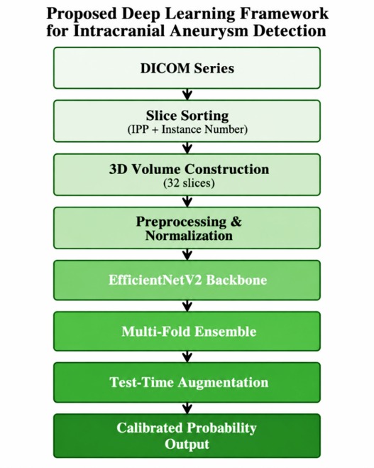
Intracranial aneurysms (IAs) have a case-fatality rate greater than 50% following rupture, and the majority are clinically silent until hemorrhage occurs. An important research direction in neurovascular imaging informatics is the development of automated screening tools that can identify potentially high-risk cases for expedited expert review. In this work, we propose a binary deep learning framework for patient-level aneurysm detection from volumetric DICOM neuroimaging. The proposed architecture adapts EfficientNetV2-S to a 2.5D input representation of channel-stacked 32 axial slices resized to 384 × 384 pixels. Percentile-based intensity normalization is employed to reduce cross-scanner variability. We measure model generalization using stratified five-fold cross-validation and improve prediction stability using weighted ensemble averaging, test-time augmentation, and temperature-scaled probability calibration. On the RSNA 2025 Intracranial Aneurysm Detection Challenge dataset of 1,147 clinical cases, the framework achieved a weighted AUC-ROC of 0.694 with a cross-validation mean of 0.681±0.005. These results indicate that the proposed method can be considered as a first research baseline for automated aneurysm pre-screening, but the performance obtained is still not enough to be used in the clinical routine without assistance. Before its integration into clinical radiology workflows, it needs external validation on independent multi-site cohorts, threshold-specific sensitivity and specificity analysis, and lesion-level evaluation when annotations are available.
Downloads
References
[1] P. M. Abbate et al., “The Cerebral Arterial Wall in the Development and Growth of Intracranial Aneurysms,” Applied Sciences 2022, Vol. 12, Page 5964, vol. 12, no. 12, p. 5964, Jun. 2022, doi: 10.3390/APP12125964. DOI: https://doi.org/10.3390/app12125964
[2] I. Rautalin et al., “Global, Regional, and National Burden of Nontraumatic Subarachnoid Hemorrhage: The Global Burden of Disease Study 2021,” JAMA Neurol., vol. 82, no. 8, pp. 765–787, May 2025, doi: 10.1001/JAMANEUROL.2025.1522. DOI: https://doi.org/10.1001/jamaneurol.2025.1522
[3] A. Mohammed Almalki et al., “Role of Radiology, Laboratory Testing, Preventive Strategies,and Nursing Care in Management of Stroke,” Saudi J Med Pharm Sci, vol. 11, no. 12, pp. 1215–1220, 2025, doi: 10.36348/sjmps.2025.v11i12.012. DOI: https://doi.org/10.36348/sjmps.2025.v11i12.012
[4] S. Ursprung, “Novel multi-parametric, multi-modality imaging for the assessment of tumour biology in renal cell carcinoma,” 2022, doi: 10.17863/CAM.80124.
[5] G. Litjens et al., “A survey on deep learning in medical image analysis,” Med. Image Anal., vol. 42, pp. 60–88, Dec. 2017, doi: 10.1016/J.MEDIA.2017.07.005. DOI: https://doi.org/10.1016/j.media.2017.07.005
[6] N. Passi, M. Raj, and N. A. Shelke, “A Review on Transformer Models: Applications, Taxonomies, Open Issues and Challenges,” 2024 4th Asian Conference on Innovation in Technology, ASIANCON 2024, 2024, doi: 10.1109/ASIANCON62057.2024.10838047. DOI: https://doi.org/10.1109/ASIANCON62057.2024.10838047
[7] K. A. S. Pillai, K. P. Preena, and M. S. Nair, “Analyzing the Efficacy of Computer-Aided Detection in Cerebral Aneurysm Diagnosis Using MRI Modality: A Review,” IEEE Access, vol. 13, pp. 12468–12482, 2025, doi: 10.1109/ACCESS.2025.3530932. DOI: https://doi.org/10.1109/ACCESS.2025.3530932
[8] X. Yang, D. J. Blezek, L. T. E. Cheng, W. J. Ryan, D. F. Kallmes, and B. J. Erickson, “Computer-Aided Detection of Intracranial Aneurysms in MR Angiography,” Journal of Digital Imaging 2009 24:1, vol. 24, no. 1, pp. 86–95, Nov. 2009, doi: 10.1007/S10278-009-9254-0. DOI: https://doi.org/10.1007/s10278-009-9254-0
[9] K. He, X. Zhang, S. Ren, and J. Sun, “Deep residual learning for image recognition,” Proceedings of the IEEE Computer Society Conference on Computer Vision and Pattern Recognition, vol. 2016-December, pp. 770–778, Dec. 2016, doi: 10.1109/CVPR.2016.90. DOI: https://doi.org/10.1109/CVPR.2016.90
[10] A. Park et al., “Deep Learning-Assisted Diagnosis of Cerebral Aneurysms Using the HeadXNet Model,” JAMA Netw. Open, vol. 2, no. 6, p. e195600, Jun. 2019, doi: 10.1001/JAMANETWORKOPEN.2019.5600. DOI: https://doi.org/10.1001/jamanetworkopen.2019.5600
[11] T. Nakao et al., “Deep neural network-based computer-assisted detection of cerebral aneurysms in MR angiography,” Journal of Magnetic Resonance Imaging, vol. 47, no. 4, pp. 948–953, Apr. 2018, doi: 10.1002/JMRI.25842. DOI: https://doi.org/10.1002/jmri.25842
[12] X. Yang, D. Xia, T. Kin, and T. Igarashi, “INTRA: 3D intracranial aneurysm dataset for deep learning,” Proceedings of the IEEE Computer Society Conference on Computer Vision and Pattern Recognition, pp. 2653–2663, 2020, doi: 10.1109/CVPR42600.2020.00273. DOI: https://doi.org/10.1109/CVPR42600.2020.00273
[13] K. M. Timmins et al., “Comparing methods of detecting and segmenting unruptured intracranial aneurysms on TOF-MRAS: The ADAM challenge,” Neuroimage, vol. 238, Sep. 2021, doi: 10.1016/j.neuroimage.2021.118216. DOI: https://doi.org/10.1016/j.neuroimage.2021.118216
[14] Z. Liu et al., “Swin Transformer: Hierarchical Vision Transformer using Shifted Windows,” Proceedings of the IEEE International Conference on Computer Vision, pp. 9992–10002, 2021, doi: 10.1109/ICCV48922.2021.00986. DOI: https://doi.org/10.1109/ICCV48922.2021.00986
[15] M. Orouskhani et al., “Intracranial aneurysm segmentation with nnU-net: utilizing loss functions and automated vessel extraction,” Vessel Plus. 2025;9:24., vol. 9, p. N/A-N/A, Dec. 2025, doi: 10.20517/2574-1209.2025.42. DOI: https://doi.org/10.20517/2574-1209.2025.42
[16] D. Ueda et al., “Deep Learning for MR Angiography: Automated Detection of Cerebral Aneurysms,” https://doi.org/10.1148/radiol.2018180901, vol. 290, no. 1, pp. 187–194, Oct. 2018, doi: 10.1148/RADIOL.2018180901. DOI: https://doi.org/10.1148/radiol.2018180901
[17] F. Isensee, P. F. Jaeger, S. A. A. Kohl, J. Petersen, and K. H. Maier-Hein, “nnU-Net: a self-configuring method for deep learning-based biomedical image segmentation,” Nature Methods 2020 18:2, vol. 18, no. 2, pp. 203–211, Dec. 2020, doi: 10.1038/s41592-020-01008-z. DOI: https://doi.org/10.1038/s41592-020-01008-z
[18] M. Tan and Q. V Le, “EfficientNet: Rethinking Model Scaling for Convolutional Neural Networks,” 2019. Accessed: Jun. 02, 2026. [Online]. Available: https://mlanthology.org/icml/2019/tan2019icml-efficientnet/
[19] M. Tan and Q. V Le, “EfficientNetV2: Smaller Models and Faster Training,” Jul. 01, 2021, PMLR. Accessed: Jun. 02, 2026. [Online]. Available: https://proceedings.mlr.press/v139/tan21a.html
[20] J. Deng, W. Dong, R. Socher, L. J. Li, K. Li, and L. Fei-Fei, “ImageNet: A Large-Scale Hierarchical Image Database,” 2009 IEEE Conference on Computer Vision and Pattern Recognition, CVPR 2009, pp. 248–255, 2009, doi: 10.1109/CVPR.2009.5206848. DOI: https://doi.org/10.1109/CVPR.2009.5206848
[21] A. A. Hassanain, T. W. Hong, R. Kumar, and M. A. Abdelrahman, “Hierarchical Swin Transformer for Multi-Stage Dementia Diagnosis with Clinically-Grounded Visual Explainability,” Journal of Smart Algorithms and Applications (JSAA), vol. 3, no. 2, pp. 105–116, Apr. 2026, doi: 10.66279/j4m1km41. DOI: https://doi.org/10.66279/j4m1km41
[22] H. Saeed et al., “Reliable Drug–Target Interaction Prediction Using Convolutional Neural Networks with Robust Negative Sample Generation,” Journal of Smart Algorithms and Applications (JSAA), vol. 2, no. 2, pp. 34–48, Feb. 2026, Accessed: Jun. 26, 2026. [Online]. Available: https://pub.scientificirg.com/index.php/JSAA/article/view/47
[23] Z. Qiu, T. Yao, and T. Mei, “Learning Spatio-Temporal Representation With Pseudo-3D Residual Networks,” 2017. DOI: https://doi.org/10.1109/ICCV.2017.590
[24] O. Ronneberger, P. Fischer, and T. Brox, “U-net: Convolutional networks for biomedical image segmentation,” Lecture Notes in Computer Science (including subseries Lecture Notes in Artificial Intelligence and Lecture Notes in Bioinformatics), vol. 9351, pp. 234–241, 2015, doi: 10.1007/978-3-319-24574-4_28/SAVE-RESEARCH. DOI: https://doi.org/10.1007/978-3-319-24574-4_28
Downloads
Published
Data Availability Statement
This study is based on a collection of medical scans. These have been taken from different hospitals with various setups. Some are thin layers, and some are thick; each source is different in its scanning methods.
Issue
Section
Categories
License
Copyright (c) 2026 Journal of Smart Algorithms and Applications (JSAA)

This work is licensed under a Creative Commons Attribution 4.0 International License.
Journal of Smart Algorithms and Applications (JSAA) content is published under a Creative Commons Attribution License (CCBY). This means that content is freely available to all readers upon publication, and content is published as soon as production is complete.
Journal of Smart Algorithms and Applications (JSAA) seeks to publish the most influential papers that will significantly advance scientific understanding. Selected articles must present new and widely significant data, syntheses, or concepts. They should merit recognition by the wider scientific community and the general public through publication in a reputable scientific journal.